3D Gait Analysis System
Visualizing Movement Biomechanics
Markerless · Fully Quantified · Rapid Assessment — covering clinical rehab, research, and health management
Core Advantages
Powered by 3D Vision AI, breaking through traditional assessment limitations
Markerless Precision Capture
Markerless camera-based movement analysis provides joint-angle and biomechanical outputs; available measures and capture settings depend on the configured product and protocol.
Full-Dimension Quantification
Outputs stride length, cadence, joint angles and other spatio-temporal parameters, plus lower limb joint forces and muscle activation biomechanical metrics.
Multi-Modal Visualization
Supports synchronized video playback, parameter tables, joint angle curves, and foot trajectory maps — data precisely matched to movement.
Low Barrier to Entry
No biomechanics expertise required. Everyday clinical staff can operate quickly, needing only 2-3 meters of test space.
Feature Highlights
Covering the complete assessment workflow
Full Gait Cycle Analysis
Covers foot contact, support, extension, and swing phases. Outputs left/right mean values and ranges, precisely identifying gait abnormalities.
Multi-Dimension Output
Spatio-temporal (stride, cadence, velocity), kinematics (joint angles), dynamics (joint forces), and muscle activation — four core data categories.
Visual Data Interpretation
Tabular precise values, joint angle curves with annotated extremes, foot path trajectories for intuitive movement pattern display.
Report Generation & Export
AI auto-generates structured assessment reports. Supports print and electronic download.
Multi-Period Comparison
Supports cross-session data comparison, intuitively showing rehabilitation or training improvement trends.
Report Example
Structured report template, precisely quantifying assessment characteristics
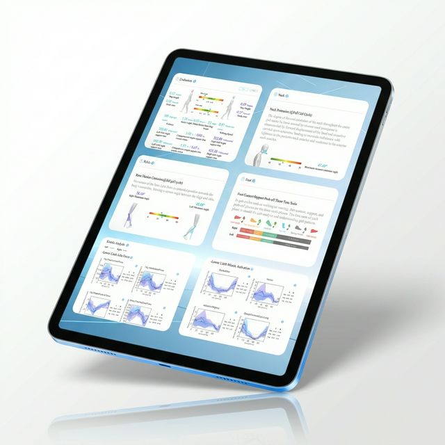
How to read and compare the report
The images on this page illustrate the HoloMotion report interface. Open the original image to inspect the layout; it is not a new accuracy experiment or evidence of treatment benefit.
Read parameter tables together with the recorded movement, side and angle convention. Check axes and units before comparing curves. A percentage displayed as a movement-risk index is not necessarily a calibrated probability of injury.
Before repeating an assessment, record product/version, task, camera position, lighting, clothing, assistance and trial-quality decisions. Compare only sufficiently similar trials and interpret change with professional context.
Existing report-interface illustration; not a complete participant report or a new validation result.
Download the interface illustration